To accommodate the increasing number of diverse blog posts by museum staff and volunteers, Ex crypta: The Curator's blog has evolved into the Museum of Health Care Blog, which can be found at http://museumofhealthcare.wordpress.com/.
All new posts, as well as the Ex crypta archives, can be found on the Museum of Health Care Blog.
Thursday, August 4, 2011
Tuesday, June 14, 2011
Thank you for your Patronage to our Hall of Honour Exhibits at Kingston General Hospital
 |
Curatorial Assistant Erin Manning (L) and Collections Intern Tanya Szulga (R) |
Over the past twenty years the Museum of Health Care has created exhibits for the Kingston General Hospital’s Hall of Honour. Recently KGH staff is working on a new redesign of this area and as part of that design process asked the museum to remove the exhibits for construction and carpet removal due to begin in July 2011.
To facilitate the dismantling of the four current exhibits, two museum studies students were engaged to lead the multi-faceted process of returning the items to the museum and loaned items, cleaning and preparing for storage all objects. Tanya Szulga, Collections Intern from Fleming College and Erin Manning, Collections Technician from Algonquin College were the perfect choice to ensure the safety and long term condition of the various items on exhibit.
Dismantling an on-site exhibit and the multiple stages of preparation and activity required for one exhibit is intense but this project involved four exhibits located off-site. Logistics and lots of preparation were required. The following provides a glimpse at what it takes to take down an exhibit following museum standards.
Dismantling the exhibits on display in the Kingston General Hospital’s Hall of Honour began weeks before the actual removal process commenced. Tanya Szulga prepared detailed exhibit artefact lists, storage location charts and supply lists to ensure that all objects would be properly transported back to the museum, had ‘homes’ in the storage areas to return to, and to ensure that the information on the database would be up-to-date and complete with pictures of all items by the end of the process. She also created a step-by-step guide to prepare for the removal process and make the days of dismantling as smooth and problem-free as possible.
Thursday, June 2nd 2011 was a perfect summer day with sunny weather forecast a great start to begin the dismantling and transportation of the first group of exhibits to be dismantled. Tanya and Erin began dismantling the “White Plaque: Fighting TB” and “Beyond Ether: Anesthesia” exhibits. The last exhibit “KGH Auxiliary” had the most quantity of items and was completed the next day.
 |
Throughout the process the pair was constantly stopped by hospital staff and visitors wishing to express how pleased they were over the years to see the items on exhibit, and now saddened to hear the new exhibits will not be returning for a possible one to two years. They expressed hope that the Kingston General Hospital could decide on the redesign of the Hall of Honour soon and allow the Museum of Health Care staff to re-install new exhibits in the future. Tanya and Erin reminded all these people that while the Hall of Honour exhibits are now gone, the Museum, located next door to Kingston General Hospital is open Tuesday to Sunday starting 18 June from 10-4 pm, and the numerous exhibits on the main level at the museum show a variety of interesting aspects of medical history. We even have a large display on the history of nurse’s uniforms and a restored nursing residence bedroom. The new Children’s Gallery has a very colourful wall mural that everyone enjoys.
An era comes to a close with the removal of this group of four exhibits and museum staff eagerly wait for the nod from KGH staff that the new design of the exhibit cases meet museum standards and new medical history topics can be researched, items selected and installed for the viewing public.
Tanya Szulga, Collections Intern
Interview with Former Curator Paul Robertson
As mentioned at the bottom of the previous blog post, after seven years with the Museum of Health Care, former Curator Paul Robertson has decided to move on. The Museum of Health Care thanks Paul for his many years of innovative, exciting curatorial work and wishes him all the best in his new position.
Before his departure, we conducted a mini interview with Paul. Here are Paul's answers:
Why did you become a curator? from MuseumOfHealthCare on Vimeo.
What's a day in the life of a curator like? from MuseumOfHealthCare on Vimeo.
How did you become a curator? from MuseumOfHealthCare on Vimeo.
We are pleased to announce that we will be featuring some guest posts from other members of our curatorial staff this summer, so please stay tuned!
Thursday, May 19, 2011
Artificial Placenta Project
 |
Mr. Jean Fortin |
Such is the case with a donation the Museum received in 2010 from Mr. Jean Fortin, a retired marketing specialist in biomedical products. Mr. Fortin’s story intrigued me when he first contacted the Museum. He wanted to offer us a small collection of papers documenting his role in the early 1960s in the development of an artificial placenta at the University of Alberta. This project was led by Dr. John C. Callaghan, a cardiac surgeon at the university hospital noted for setting up the its open heart surgery unit in 1956 and performing the first successful cardiac operations in Canada using the heart-lung pump. He had previously worked in Toronto in the early 1950s with Dr. W.G. Bigelow to develop a first-generation intravenous pacemaker, an ancestor of modern day pacing.
I found Mr. Fortin’s role in this work interesting because he was one of the technicians involved in the artificial placenta project, an occupation often underreported in historical documentation of scientific and medical advances. Born in Quebec and originally trained in the 1950s as a Royal Canadian Navy electronic technician, Mr. Fortin joined Dr. Callaghan’s team at the University of Alberta research laboratories in 1962. By 1964, he was responsible for maintaining all of the electronic equipment for the artificial placenta project.
The placenta machine was designed to nurture a lamb removed by caesarean section from its mother. Before it could breathe on its own, it was placed in a plastic temperature-controlled bowl or artificial womb filled with a solution of sugar and salts replicating amniotic fluid. The lamb could be kept alive with a newly developed heart-lung machine that performed the oxygenating function of the placenta, the organ through which all unborn mammals receive oxygen and food from their mothers. Because they have similar physiological needs to human babies, lambs were often used during this period as stand-ins for humans in experiments.
The experiment’s main purpose was to devise ways to improve heart-lung machines that had already become standard in many hospitals. Although essential equipment for surgeons performing open heart surgery, existing heart-lung machines tended to damage blood cells after five or six hours of continuous use and therefore were used mainly for surgery and never as a life-saving aid for a baby born prematurely. Dr. Callaghan’s intention was to perfect a heart-lung machine that oxygenated blood so gently that premature babies with lung ailments could be kept alive until they were able to breathe on their own.
Mr. Fortin’s responsibilities including tending a flock of 200 ewes and one ram to provide the project with lambs, but more significantly, he was the technician who developed the artificial lung component of the artificial placenta unit. As he explained to me, that portion of the machine was missing until 1964 when he managed to adapt an artificial kidney design to act as a ‘lung’ oxygenator.
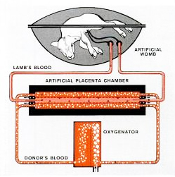 |
Image source: Life Magazine |
In August 1964 the research team had its first successful experiment when one lamb survived for 21 hours in the machine and lived when it was removed. Mr. Fortin reports this lamb was named John Glenn after the first American astronaut to orbit the earth. The project received national coverage in that month’s edition of Life magazine (which also featured a cover story on the Beatles).
Mr. Fortin later became a regular member of the university hospital’s cardiac perfusion team running the heart-lung pump during open heart surgeries. By 1966 he had assisted in 500 operations. He told me of one dramatic incident during an open heart operation when the pump that provided the oxygenated arterial blood seized five minutes into the operation. Mr. Fortin was called out to operate the machine manually by turning a crank for 90 minutes until the surgery was completed. The patient survived.
Mr. Fortin later left the university to go into sales of cardiac monitoring equipment. He retired in 2005. We are grateful to Mr. Fortin for bringing this personal story of health care history in Canada to the Museum.
Paul Robertson
Note: After seven years with the Museum of Health Care Paul Robertson has decided to move on. The Museum of Health Care thanks Paul for his many years of innovative, exciting curatorial work and wishes him all the best in his new position. Although this will be Paul’s final blog post for Ex Crypta, the blog may continue in the future… stay tuned!
Subscribe to:
Posts (Atom)
About Me

- Museum of Health Care Staff
- The Museum of Health Care shows how Canadians have preserved health and managed disease, pain and suffering. The Museum strives to connect visitors of all ages with the experience of people in past times and provide context and perspective on today's health issues.