To accommodate the increasing number of diverse blog posts by museum staff and volunteers, Ex crypta: The Curator's blog has evolved into the Museum of Health Care Blog, which can be found at http://museumofhealthcare.wordpress.com/.
All new posts, as well as the Ex crypta archives, can be found on the Museum of Health Care Blog.
Thursday, August 4, 2011
Tuesday, June 14, 2011
Thank you for your Patronage to our Hall of Honour Exhibits at Kingston General Hospital
 |
Curatorial Assistant Erin Manning (L) and Collections Intern Tanya Szulga (R) |
Over the past twenty years the Museum of Health Care has created exhibits for the Kingston General Hospital’s Hall of Honour. Recently KGH staff is working on a new redesign of this area and as part of that design process asked the museum to remove the exhibits for construction and carpet removal due to begin in July 2011.
To facilitate the dismantling of the four current exhibits, two museum studies students were engaged to lead the multi-faceted process of returning the items to the museum and loaned items, cleaning and preparing for storage all objects. Tanya Szulga, Collections Intern from Fleming College and Erin Manning, Collections Technician from Algonquin College were the perfect choice to ensure the safety and long term condition of the various items on exhibit.
Dismantling an on-site exhibit and the multiple stages of preparation and activity required for one exhibit is intense but this project involved four exhibits located off-site. Logistics and lots of preparation were required. The following provides a glimpse at what it takes to take down an exhibit following museum standards.
Dismantling the exhibits on display in the Kingston General Hospital’s Hall of Honour began weeks before the actual removal process commenced. Tanya Szulga prepared detailed exhibit artefact lists, storage location charts and supply lists to ensure that all objects would be properly transported back to the museum, had ‘homes’ in the storage areas to return to, and to ensure that the information on the database would be up-to-date and complete with pictures of all items by the end of the process. She also created a step-by-step guide to prepare for the removal process and make the days of dismantling as smooth and problem-free as possible.
Thursday, June 2nd 2011 was a perfect summer day with sunny weather forecast a great start to begin the dismantling and transportation of the first group of exhibits to be dismantled. Tanya and Erin began dismantling the “White Plaque: Fighting TB” and “Beyond Ether: Anesthesia” exhibits. The last exhibit “KGH Auxiliary” had the most quantity of items and was completed the next day.
 |
Throughout the process the pair was constantly stopped by hospital staff and visitors wishing to express how pleased they were over the years to see the items on exhibit, and now saddened to hear the new exhibits will not be returning for a possible one to two years. They expressed hope that the Kingston General Hospital could decide on the redesign of the Hall of Honour soon and allow the Museum of Health Care staff to re-install new exhibits in the future. Tanya and Erin reminded all these people that while the Hall of Honour exhibits are now gone, the Museum, located next door to Kingston General Hospital is open Tuesday to Sunday starting 18 June from 10-4 pm, and the numerous exhibits on the main level at the museum show a variety of interesting aspects of medical history. We even have a large display on the history of nurse’s uniforms and a restored nursing residence bedroom. The new Children’s Gallery has a very colourful wall mural that everyone enjoys.
An era comes to a close with the removal of this group of four exhibits and museum staff eagerly wait for the nod from KGH staff that the new design of the exhibit cases meet museum standards and new medical history topics can be researched, items selected and installed for the viewing public.
Tanya Szulga, Collections Intern
Interview with Former Curator Paul Robertson
As mentioned at the bottom of the previous blog post, after seven years with the Museum of Health Care, former Curator Paul Robertson has decided to move on. The Museum of Health Care thanks Paul for his many years of innovative, exciting curatorial work and wishes him all the best in his new position.
Before his departure, we conducted a mini interview with Paul. Here are Paul's answers:
Why did you become a curator? from MuseumOfHealthCare on Vimeo.
What's a day in the life of a curator like? from MuseumOfHealthCare on Vimeo.
How did you become a curator? from MuseumOfHealthCare on Vimeo.
We are pleased to announce that we will be featuring some guest posts from other members of our curatorial staff this summer, so please stay tuned!
Thursday, May 19, 2011
Artificial Placenta Project
 |
Mr. Jean Fortin |
Such is the case with a donation the Museum received in 2010 from Mr. Jean Fortin, a retired marketing specialist in biomedical products. Mr. Fortin’s story intrigued me when he first contacted the Museum. He wanted to offer us a small collection of papers documenting his role in the early 1960s in the development of an artificial placenta at the University of Alberta. This project was led by Dr. John C. Callaghan, a cardiac surgeon at the university hospital noted for setting up the its open heart surgery unit in 1956 and performing the first successful cardiac operations in Canada using the heart-lung pump. He had previously worked in Toronto in the early 1950s with Dr. W.G. Bigelow to develop a first-generation intravenous pacemaker, an ancestor of modern day pacing.
I found Mr. Fortin’s role in this work interesting because he was one of the technicians involved in the artificial placenta project, an occupation often underreported in historical documentation of scientific and medical advances. Born in Quebec and originally trained in the 1950s as a Royal Canadian Navy electronic technician, Mr. Fortin joined Dr. Callaghan’s team at the University of Alberta research laboratories in 1962. By 1964, he was responsible for maintaining all of the electronic equipment for the artificial placenta project.
The placenta machine was designed to nurture a lamb removed by caesarean section from its mother. Before it could breathe on its own, it was placed in a plastic temperature-controlled bowl or artificial womb filled with a solution of sugar and salts replicating amniotic fluid. The lamb could be kept alive with a newly developed heart-lung machine that performed the oxygenating function of the placenta, the organ through which all unborn mammals receive oxygen and food from their mothers. Because they have similar physiological needs to human babies, lambs were often used during this period as stand-ins for humans in experiments.
The experiment’s main purpose was to devise ways to improve heart-lung machines that had already become standard in many hospitals. Although essential equipment for surgeons performing open heart surgery, existing heart-lung machines tended to damage blood cells after five or six hours of continuous use and therefore were used mainly for surgery and never as a life-saving aid for a baby born prematurely. Dr. Callaghan’s intention was to perfect a heart-lung machine that oxygenated blood so gently that premature babies with lung ailments could be kept alive until they were able to breathe on their own.
Mr. Fortin’s responsibilities including tending a flock of 200 ewes and one ram to provide the project with lambs, but more significantly, he was the technician who developed the artificial lung component of the artificial placenta unit. As he explained to me, that portion of the machine was missing until 1964 when he managed to adapt an artificial kidney design to act as a ‘lung’ oxygenator.
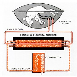 |
Image source: Life Magazine |
In August 1964 the research team had its first successful experiment when one lamb survived for 21 hours in the machine and lived when it was removed. Mr. Fortin reports this lamb was named John Glenn after the first American astronaut to orbit the earth. The project received national coverage in that month’s edition of Life magazine (which also featured a cover story on the Beatles).
Mr. Fortin later became a regular member of the university hospital’s cardiac perfusion team running the heart-lung pump during open heart surgeries. By 1966 he had assisted in 500 operations. He told me of one dramatic incident during an open heart operation when the pump that provided the oxygenated arterial blood seized five minutes into the operation. Mr. Fortin was called out to operate the machine manually by turning a crank for 90 minutes until the surgery was completed. The patient survived.
Mr. Fortin later left the university to go into sales of cardiac monitoring equipment. He retired in 2005. We are grateful to Mr. Fortin for bringing this personal story of health care history in Canada to the Museum.
Paul Robertson
Note: After seven years with the Museum of Health Care Paul Robertson has decided to move on. The Museum of Health Care thanks Paul for his many years of innovative, exciting curatorial work and wishes him all the best in his new position. Although this will be Paul’s final blog post for Ex Crypta, the blog may continue in the future… stay tuned!
Thursday, September 23, 2010
Fenwick Operating Theatre: a life-saving surgery in Edwardian Kingston
 |
Bennie Stalker two weeks after surgery, October 1901, Source: Jim Bremner |
These events occurred in eastern Ontario in September - October 1901. This account reveals much about the stark realities of rural Canadian health care a century ago, but at the same time, the amazing ability of the human body to survive severe trauma and the abiding human desire to care for the sick.
At the heart of the story is young William Benjamin Stalker, who was born in 1891 and lived on a farm near Plevna. The clinical details of his misadventure and life-saving surgery are preserved in the surgeon’s report detailing the boy’s accident and medical treatment in the January 1902 Kingston Medical Quarterly. For historians, Dr. W.G. Anglin’s case study puts a human face to the ancient hospital spaces and dry administrative reports that remain as historical evidence today.
After Bennie’s all-day journey to Kingston General Hospital, he was admitted to the St. Andrew’s Ward for children in the upper storey of the Watkins Wing. Dr. Anglin describes in detail the severity of the boy’s wounds and the extreme deterioration of his arm and upper extremity. The next day we learn that Bennie was wheeled into the recently constructed Fenwick Operating Theatre. Complete amputation was the only option if the boy was to survive – grim, but effective treatment. News reports of the emergency amputations required to save lives of victims of the January 2010 earthquake in Haiti spring to mind.
 |
Gigli saw for cutting bone, MHC Collection 1976.6.37 |
The doctor’s description of Bennie’s aftercare says much about social services of the day: “Our readers may be interested to know that having but one parent living, the little fellow has been admitted into that excellent institution the Orphans’ Home in this city.”
Dr. Anglin recorded that Bennie was an interesting patient and always quick and bright with his answers. He recounted the boy’s encounter with Dr. Alan Manby, the physician accompanying the Duke and Duchess of Cornwall and York (King George V and Queen Mary) while on their 1901 cross-Canada tour. During a visit to KGH Dr. Manby told Bennie that he would be unable to visit him again because of the great distance the doctor had come to be there. Bennie’s quick and unexpected response is reported to be, “”Well, I came nearly a hundred miles myself to get here.” How medical journal articles have changed.
After his amazing recovery, Bennie grew up to have a full and productive life, first as a travelling ventriloquist and later an itinerant photographer. He married and had five children. After Bennie’s death in 1940, his wife remarried and had a son named Jim Bremner. I thank Mr. Bremner and his wife Marianne who spent ten years researching Bennie’s fascinating story for bringing it to our attention. This is what helps to make history real and bring museum collections to life.
Paul Robertson
Curator
Tuesday, April 20, 2010
Funding Success for Museum Collection
 |
Iron lung, built at Toronto's Hospital for Sick Children, 1937, MHC Collection 997.019.003 |
Although most of this material is currently available for viewing on our publicly accessible online collections catalogue, we know that many of our most intriguing items are largely hidden from the average visitor to the Museum’s website, particularly from people who may not feel comfortable navigating a database. Another disadvantage is that the artefacts in the catalogue are not searchable from outside the website. It will now be possible to search for objects featured in the new artefact profiles with Google and other external search engines.
 |
Enema Syringe, circa 1800, MHC Collection 002.050.006 a-d |
We want to involve the public more with our collection. “From the Collection” will include a link to a newly created online forum included with each profile. In this way we hope to provide an opportunity for user-generated content through community contribution, discussion, and interaction.
Watch this site for further information as the project gets under way!
Paul Robertson
Curator
Friday, February 19, 2010
Early Penicillin Sample Comes to Collection
Curators are always excited when they make a “find”, especially when that find more or less just arrives at our doorstep: an ampoule containing some of the first experimental penicillin produced in Canada!
How did it come to the Museum of Health Care? In the late 1990s the Museum received a transfer of artefacts and archival documents from Queen’s University (Kingston, Canada), known as the Faculty of Medicine Collection. Most of the objects were shifted to the Museum at that time, but a few remained on display in the medical school on campus. Recently it was decided to move the remaining pieces to the Museum for processing and preservation. Among the Victorian surgeon’s kits, textbooks, and medical student graduation programmes was a nine centimetre glass vial holding a white powder and a typewritten file card:
“The last of twelve ampoules containing the first batch of PENICILLIN (10,000 units) made experimentally by Ayerst, McKenna, Harrison of Montreal. The untried, unproved drug was used successfully (but unofficially) to save the life of a 16 year old boy, critically ill with septicemia following a ruptured appendix in the summer of 1940.”
According to the card, the sample was given to Queen’s by one of its grads, Dr. C.W. Kelley (1928), former chief of surgery, Ottawa Civic Hospital.
The anti-bacterial function of penicillin was first discovered in 1928 by English bacteriologist Alexander Fleming, but its clinical potential was not realised until 1940 when pathologist Howard Florey and biochemist Ernst Chain were able to extract, purify, and produce the drug in their laboratory. This short video explains the discovery of penicillin:
What makes our penicillin sample particularly rare is that the isolation of the antibiotic’s active ingredient in England appears to have taken place only a short while before this ampoule was produced. Founded in 1925, Ayerst, McKenna and Harrison was a young Canadian pharmaceutical firm – in 1931 it set up the first commercial biological laboratory in the country. Penicillin was still an experimental drug and clearly its use on a teenager in 1940 was a gamble, but fortunately one with a happy outcome, given how unauthorised was its administration.
During the Second World War pharmaceutical companies in several countries rushed to produce penicillin for soldiers – the antibiotic’s ability to greatly reduce mortality rates resulting from infected wounds, unclean surgery, and infectious diseases was a clear advantage on the battlefield. The war galvanised the mass production of many drugs and penicillin became available on a wide scale to the general public between 1944 and 1946. This 20th –century “magic bullet” has long been an effective weapon against pneumonia, anthrax, tetanus, syphilis, and diphtheria. The little penicillin ampoule buried in a university exhibit showcase – what has turned out to be an important treasure highlighting a significant development in Canadian healthcare history.
Paul Robertson
Curator
 |
Ampoule of penicillin, MHC Collection |
“The last of twelve ampoules containing the first batch of PENICILLIN (10,000 units) made experimentally by Ayerst, McKenna, Harrison of Montreal. The untried, unproved drug was used successfully (but unofficially) to save the life of a 16 year old boy, critically ill with septicemia following a ruptured appendix in the summer of 1940.”
According to the card, the sample was given to Queen’s by one of its grads, Dr. C.W. Kelley (1928), former chief of surgery, Ottawa Civic Hospital.
The anti-bacterial function of penicillin was first discovered in 1928 by English bacteriologist Alexander Fleming, but its clinical potential was not realised until 1940 when pathologist Howard Florey and biochemist Ernst Chain were able to extract, purify, and produce the drug in their laboratory. This short video explains the discovery of penicillin:
What makes our penicillin sample particularly rare is that the isolation of the antibiotic’s active ingredient in England appears to have taken place only a short while before this ampoule was produced. Founded in 1925, Ayerst, McKenna and Harrison was a young Canadian pharmaceutical firm – in 1931 it set up the first commercial biological laboratory in the country. Penicillin was still an experimental drug and clearly its use on a teenager in 1940 was a gamble, but fortunately one with a happy outcome, given how unauthorised was its administration.
During the Second World War pharmaceutical companies in several countries rushed to produce penicillin for soldiers – the antibiotic’s ability to greatly reduce mortality rates resulting from infected wounds, unclean surgery, and infectious diseases was a clear advantage on the battlefield. The war galvanised the mass production of many drugs and penicillin became available on a wide scale to the general public between 1944 and 1946. This 20th –century “magic bullet” has long been an effective weapon against pneumonia, anthrax, tetanus, syphilis, and diphtheria. The little penicillin ampoule buried in a university exhibit showcase – what has turned out to be an important treasure highlighting a significant development in Canadian healthcare history.
Paul Robertson
Curator
Subscribe to:
Posts (Atom)
About Me

- Museum of Health Care Staff
- The Museum of Health Care shows how Canadians have preserved health and managed disease, pain and suffering. The Museum strives to connect visitors of all ages with the experience of people in past times and provide context and perspective on today's health issues.